


Traditional body mass index (BMI) calculations have long served as the primary metric for assessing weight-related health risks, yet this simple height-to-weight ratio fails to distinguish between muscle mass, bone density, and adipose tissue distribution. For healthcare practitioners seeking precise body composition analysis, medical imaging techniques offer unprecedented accuracy in evaluating fat distribution, muscle mass, and metabolic health indicators that BMI cannot detect.
What Are the Primary Medical Imaging Techniques for Body Composition Analysis?
Modern medical imaging encompasses several sophisticated modalities specifically designed for comprehensive body composition evaluation. Each technique offers distinct advantages depending on clinical requirements, patient characteristics, and diagnostic objectives.
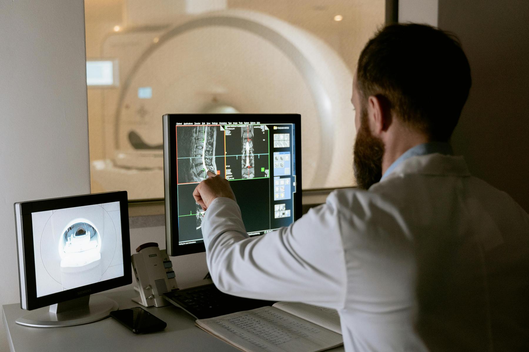
Dual-Energy X-ray Absorptiometry (DEXA) represents the current gold standard for clinical body composition assessment. This technique utilises two distinct X-ray energy levels to differentiate between bone mineral, lean tissue, and adipose tissue with exceptional precision. DEXA scanning provides whole-body and regional analysis, measuring bone mineral density simultaneously with soft tissue composition.
Magnetic Resonance Imaging (MRI) offers unparalleled soft tissue contrast resolution, enabling detailed visualisation of visceral and subcutaneous fat compartments. MRI-based body composition analysis excels in differentiating between various adipose tissue depots and provides volumetric measurements of organ-specific fat accumulation, particularly valuable for hepatic steatosis assessment.
Computed Tomography (CT) delivers cross-sectional imaging with excellent spatial resolution for adipose tissue quantification. CT scanning proves particularly effective for measuring visceral adipose tissue area and density, providing clinically relevant data for cardiovascular risk stratification.
Bioelectrical Impedance Analysis (BIA), whilst not strictly a medical imaging technique, deserves mention as a complementary assessment tool. BIA measures electrical conductivity differences between body tissues, offering rapid body composition estimates that correlate reasonably well with imaging-based measurements.
How Does DEXA Scanning Revolutionise Body Fat Assessment?
DEXA scanning has transformed clinical body composition analysis through its unique ability to provide precise, reproducible measurements across three distinct tissue compartments: bone mineral content, lean soft tissue, and adipose tissue. The scanning process involves minimal radiation exposure—approximately equivalent to one day of natural background radiation—making it suitable for repeated monitoring in clinical populations. DEXA systems generate detailed body composition reports that include total body fat percentage, regional fat distribution, lean muscle mass quantification, and bone mineral density measurements.
Clinical Applications of DEXA Scanning:
DEXA’s precision enables detection of subtle changes in body composition that traditional methods might miss. The technique proves invaluable for monitoring treatment responses in weight management programmes, where distinguishing between fat loss and muscle preservation becomes crucial for optimising outcomes.
Limitations and Considerations:
Despite its clinical utility, DEXA scanning presents certain limitations. The technique assumes standard hydration levels and may be influenced by recent food intake, exercise, or medication use. Additionally, DEXA cannot distinguish between visceral and subcutaneous abdominal fat—a distinction crucial for metabolic risk assessment.
When Should MRI and CT Scanning Be Used for Body Composition Analysis?
MRI and CT scanning serve specialised roles in body composition assessment, particularly when detailed visceral fat analysis or organ-specific fat quantification becomes clinically necessary. These modalities offer superior spatial resolution and tissue discrimination compared to DEXA scanning.
MRI Applications in Body Composition:
MRI excels in measuring ectopic fat deposition—abnormal fat accumulation within organs such as the liver, pancreas, and skeletal muscle. This capability proves essential for assessing non-alcoholic fatty liver disease, pancreatic fat infiltration, and intramyocellular lipid content, all significant metabolic health indicators.
CT Scanning Advantages:
CT imaging offers rapid acquisition times and excellent spatial resolution for visceral adipose tissue quantification. Single-slice CT measurements at the L4-L5 vertebral level correlate strongly with total visceral fat volume, providing clinically relevant data for cardiovascular risk assessment.
Clinical Decision-Making:
The choice between MRI and CT depends on several factors including radiation exposure considerations, patient contraindications, imaging time requirements, and specific clinical questions. MRI avoids ionising radiation exposure but requires longer scanning times and may be contraindicated in patients with certain metallic implants.
Why Are Medical Imaging Techniques Superior to Traditional Methods?
Medical imaging techniques provide quantitative, objective measurements that surpass traditional anthropometric assessments in both accuracy and clinical relevance. The superiority stems from their ability to directly visualise and quantify internal body composition rather than relying on indirect estimations.
Precision and Reproducibility:
Imaging-based measurements demonstrate significantly lower measurement error compared to skinfold thickness assessments, bioelectrical impedance, or BMI calculations. DEXA scanning, for instance, shows coefficient of variation values typically below 2% for repeat measurements, ensuring reliable monitoring of composition changes over time.
Clinical Relevance:
Unlike BMI, which cannot distinguish between muscle and fat mass, medical imaging techniques provide detailed tissue-specific information. This distinction proves crucial for athletes with high muscle mass, elderly individuals experiencing sarcopenia, or patients undergoing weight management interventions where muscle preservation is paramount.
Metabolic Risk Assessment:
Medical imaging enables direct measurement of visceral adipose tissue, the fat depot most strongly associated with metabolic complications. Traditional measurements cannot reliably assess this critical risk factor, potentially missing significant metabolic abnormalities in individuals with normal BMI but elevated visceral fat.
| Assessment Method | Accuracy Level | Tissue Differentiation | Radiation Exposure | Cost Consideration | Time Required |
|---|---|---|---|---|---|
| DEXA Scanning | High | Bone/Lean/Fat | Minimal | Moderate | 10-20 minutes |
| MRI | Very High | Excellent | None | High | 30-60 minutes |
| CT Scanning | High | Good | Moderate | High | 5-15 minutes |
| BIA | Moderate | Limited | None | Low | 2-5 minutes |
| BMI | Low | None | None | None | 1 minute |
Which Technique Is Most Appropriate for Different Clinical Scenarios?
Selecting appropriate body composition assessment techniques requires careful consideration of clinical objectives, patient characteristics, available resources, and diagnostic requirements. Each modality serves specific clinical niches where its particular strengths provide optimal value.
Routine Clinical Assessment:
For general clinical practice requiring comprehensive body composition analysis, DEXA scanning represents the optimal balance between accuracy, cost-effectiveness, and clinical utility. Its ability to provide bone density measurements simultaneously with body composition data offers additional diagnostic value for osteoporosis screening.
Research Applications:
Research protocols demanding maximum precision often utilise MRI for detailed fat compartment analysis or multi-compartment models combining several techniques. CT scanning serves research requiring rapid throughput or specific visceral fat measurements.
Specialised Clinical Scenarios:
Hepatology practices benefit from MRI’s capability to quantify hepatic fat content, whilst endocrinology services may prefer DEXA’s precision for monitoring sarcopenia or treatment responses in metabolic disorders. Sports medicine applications often require DEXA’s ability to distinguish between muscle and fat changes.
Population-Specific Considerations:
Paediatric populations typically receive DEXA scanning due to its minimal radiation exposure, whilst pregnant women require techniques avoiding ionising radiation entirely. Elderly patients may benefit from DEXA’s comprehensive assessment including bone density evaluation.
The Future of Medical Body Composition Assessment
Advanced medical imaging techniques continue evolving, with emerging technologies promising even greater precision and clinical utility. Artificial intelligence integration enhances measurement automation and reduces operator-dependent variability, whilst novel imaging biomarkers provide deeper insights into metabolic health status.
The integration of body composition data with other health metrics creates comprehensive patient profiles that guide personalised therapeutic approaches. This holistic assessment methodology represents a significant advancement beyond traditional weight-focused interventions, enabling more targeted and effective treatment strategies.
Medical imaging techniques for body composition assessment have fundamentally transformed our understanding of obesity, metabolic health, and treatment monitoring. As healthcare moves toward precision medicine approaches, these sophisticated assessment tools will continue playing increasingly important roles in optimising patient outcomes through evidence-based therapeutic interventions.
How accurate are DEXA scans compared to other body composition methods?
DEXA scanning demonstrates superior accuracy with measurement errors typically below 2%, significantly outperforming bioelectrical impedance analysis and anthropometric measurements. Research consistently shows DEXA provides the most reliable clinical assessment for tracking body composition changes over time.
Is radiation exposure from DEXA scans a significant health concern?
DEXA scanning involves minimal radiation exposure, approximately equivalent to one day of natural background radiation or a cross-country flight. This low exposure level makes DEXA suitable for repeated monitoring in clinical populations without significant health risks.
Can medical imaging detect visceral fat that BMI measurements miss?
Yes, medical imaging techniques, particularly MRI and CT scanning, directly measure visceral adipose tissue that BMI cannot detect. This is clinically valuable for identifying metabolic and cardiovascular risks in individuals with normal BMI but elevated visceral fat.
How often should body composition imaging be performed for monitoring purposes?
Monitoring frequency depends on clinical objectives and intervention types. Weight management programmes typically benefit from quarterly DEXA scans to track fat loss and muscle preservation, while research protocols may require more frequent assessments to detect subtle changes.
What preparation is required before medical imaging body composition assessments?
Preparation may vary by technique but generally includes avoiding intense exercise for 24 hours, maintaining normal hydration, and fasting for several hours prior to the scan to ensure accurate measurements. Specific protocols should be followed as recommended by the imaging facility.

